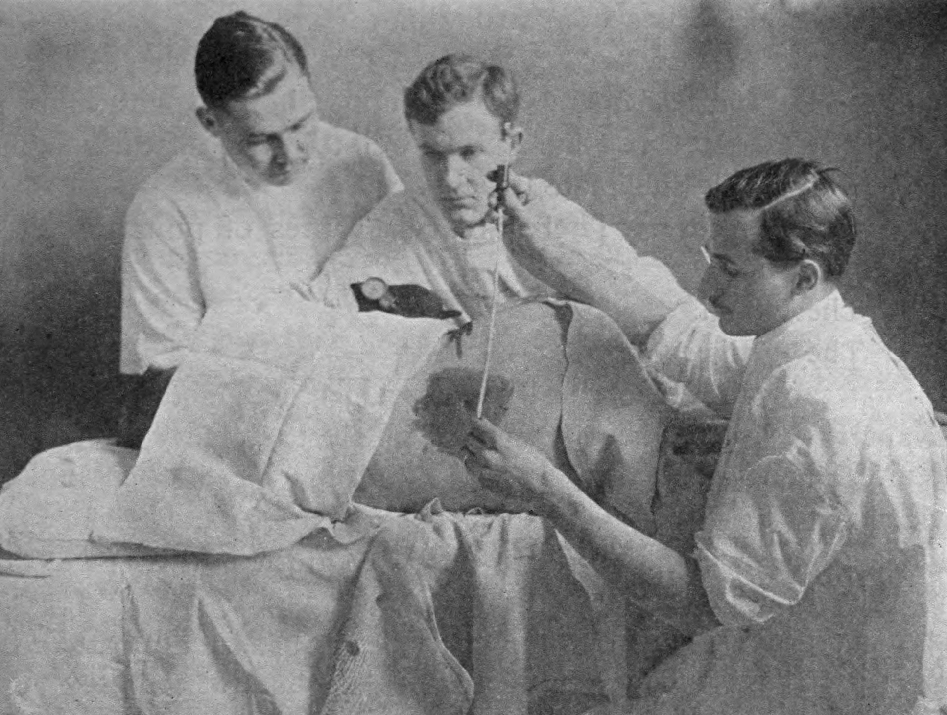
The CT-LP (lumbar puncture) diagnostic pathway has been a permanent fixture in the arsenal of the Emergency Physician for what seems like an eternity. Steadfast in its dependability, for many generations, the LP was a necessity for Emergency Physicians to safely exclude the diagnosis of subarachnoid hemorrhage (SAH). And yet, rarely a moment has passed over the past few years when Dr. Jeffrey Perry has not politely demonstrated how little we truly know about this disease process and the diagnostic tools associated with it. His 2011 paper questioning the necessity of an LP following a negative head CT under 6-hours from symptom onset, shook the once solid ground that the LP firmly stood upon (1). As if this attack on our reliable comrade was not enough, his most recent publication examining the diagnostic capabilities of the lumbar puncture itself has left our confidence in this once dependable testing strategy in turmoil.
In this paper, published in February of 2015 in The BMJ, Perry et al utilized a subset of patients from two cohorts originally enrolled to derive and validate his Ottawa SAH rule (3,4). Authors examined 1739 of these patients who received a lumbar puncture as part of their workup for SAH (2). They then sought to assess the diagnostic accuracy of this tool. Similar to common practice, they prospectively defined a positive tap as greater than 1 RBC on fluid aspirate. When this impossibly low threshold was upheld, LP's performance was less than stellar. Of the 1739 patients who received an LP, 641 (36.9%) had positive findings, only 15 of which were actually from subarachnoid blood. Most of these false positive results were trivial, as 476 (74.3%) had counts of ?100×106/L and 94 (14.8%) had counts of 101-1000×106/L. Only 10.4% of these patients were found to have clinically concerning levels of RBCs in their CSF (counts of >1000×106/L). Despite the predominance of low RBC counts, a great majority of the patients in whom the LP was positive (419) received invasive angiographic imaging.
When the LP was found to be negative (No RBCs in the CSF), it boasted a sensitivity of 100%. In an attempt to compensate for the unacceptably high number of false positives the authors retrospectively determined the ideal RBC cutoff to be 2000×106/L. At this threshold the LP had a sensitivity of 93.3% (95% confidence interval 66.0% to 99.7%) and specificity of 92.8% (90.5% to 94.6%) for aneurysmal subarachnoid hemorrhage. If visual xanthochromia was added to this RBC cutoff, the sensitivity for ruling out SAH became 100% (95% confidence interval 74.7% to 100.0%).
These numbers are of course fraught with methodological pitfalls. The threshold of 2000×106/L was retrospectively derived to best fit this specific cohort. Only 15 of the 1739 patients examined actually had the disease in question making these calculations incredibly unstable (the confidence intervals surrounding their 100% sensitivity dropped as low as 74.7%). The threshold of 2000×106/L is hardly robust enough for clinical use and will inevitably fail when applied in prospective fashion to a novel cohort.
Though this data is not definitive and further studies validating these findings are required, a number of valuable conclusions can be inferred. Surprisingly the most important of these has little to do with the diagnostic utility of the lumbar puncture.
In 2011 Perry et al published their game changing article in The BMJ examining the accuracy of a non-contrast head CT performed under 6-hours from symptom onset for the diagnosis of SAH (1). This paper was a secondary analysis of the original cohort used to derive the Ottawa SAH Rules (4). Using this preexisting cohort they assessed the accuracy of head CT for the diagnosis of SAH before and after a 6-hour threshold. The authors claim a sensitivity of 100% when CT was performed within 6-hours of symptom onset. However when the CT was performed after this 6-hour threshold, the sensitivity fell to 85.7%. Suggesting that when performed within 6-hours, a non-contrast CT is sufficient to rule out SAH, allowing practitioners to forego a subsequent lumbar puncture. Though many have viewed this as practice changing, others argue a number of flaws in the study’s design prevent us from interpreting these conclusions with such conviction.
The most obvious and often discussed weakness of this study is the use of a surrogate endpoint in place of a true gold standard. Not all patients who had a negative head CT underwent a confirmatory lumbar puncture. In its place, the authors used a 6-month proxy outcome to demonstrate the safety of CT alone. Patients underwent a structured phone interview at the 6-month mark to ascertain their wellbeing. When attempts to reach patients over the phone failed, authors endeavored to determine their status by searching medical records from regional neurosurgical centers as well as coroner’s death records. Patients were considered to be free of SAH if on 6-month follow-up they were alive and well. In the case of patients who were discovered to have passed away during the follow-up period, if the cause of death was determined to be due to something other than SAH, their deaths were not counted as a missed diagnosis. Of the 1931 patients examined, 421 were lost to follow-up. Authors found 8 of these patients had passed away since their initial workup for subarachnoid hemorrhage. Although none of these patients were determined to have died because of SAH, the reliability of post mortem cause of death is questionable at best (5).
A far less discussed aspect to this study was how the authors’ definition of a positive CT influenced the validity of their results. The standard that Perry et al used to calculate the sensitivity of head CT was based upon the Neuroradiologist’s official report. In most facilities (as was the case at the centers participating in this study) what guides Emergency Physicians’ clinical decision-making is the initial wet read usually done by Radiology house staff or even the ED physicians themselves. The sensitivity we are concerned with is that of the wet read. The Neuroradiologists in this study were not blinded to the patients’ lab findings. As such we are unable ascertain how many CTs done within 6 hours were initially read as negative, and only later after a positive LP was performed was the final report recorded as positive. If this had occurred with any frequency it would obviously harm the internal validity of the results. We are able to get a sense of how frequently this occurred by examining how many of the patients who were diagnosed with SAH had both a positive CT and LP. At least in theory, if the CT was positive then there would be no reason to perform the subsequent LP.
Of the 15 patients with SAHs that were diagnosed using a positive LP, 10 underwent head CTs and LPs that were both positive. The vast majority of these subarachnoid bleeds (n=8) were found in patients who received their CTs beyond the 6-hour threshold. There were however two patients that were identified as having received their CTs within 6-hours of symptom onset. In both these patients their initial CT was read as negative and only after a positive lumbar puncture was the final report changed to positive. If these two patients are taken into account, the adjusted sensitivity of CT under 6-hours from symptom onset is only 98.3% (with the confidence interval dropping as low as 93.6%).
These findings of course do nothing accept muddy the already cloudy waters. Head CT though fairly sensitive, will on occasion miss a subarachnoid bleed. The addition of CSF aspirate will very often offer a further degree of ambiguity. Furthermore the utilization of LP, at least in its current strategy, leads to an unacceptable number of false positives, exposing a large number of patients to needless downstream testing. If a more liberal view towards RBCs in the CSF is taken, the LP’s utility may be justifiable. Even with the retrospective best fit diagnostic capabilities calculated by Perry et al, the prevalence of SAH following a negative CT in under 6-hours is so low that further testing will likely lead to identifying far more false positive results than true subarachnoid bleeds. Cleary the conviction and certainty we once held for this testing strategy has suffered. Perhaps it is time for a shared decision making model. After all it is our patients’ value systems rather than our own biases that should guide these investigative journeys. Dr. Perry has demonstrated that the CT-LP pathway is far from straightforward. Perhaps it is time we confess these imperfections to the world at large and begin a far more honest conversation.
Sources Cited:
- Perry JJ, Stiell IG, Sivilotti ML, et al. Sensitivity of computed tomography performed within six hours of onset of headache for diagnosis of subarachnoid haemorrhage: prospective cohort study. BMJ. 2011;343:d4277.
- Perry JJ, Alyahya B, Sivilotti ML, et al. Differentiation between traumatic tap and aneurysmal subarachnoid hemorrhage: prospective cohort study. BMJ. 2015;350:h568.
- Perry JJ, Stiell IG, Sivilotti ML, et al. Clinical decision rules to rule out subarachnoid hemorrhage for acute headache. JAMA. 2013;310:(12)1248-55.
- Perry JJ, Stiell IG, Sivilotti ML, et al. High-risk clinical characteristics for subarachnoid haemorrhage in patients with acute headache: prospective cohort study. BMJ. 2010;341:c5204.
- Wexelman, BA et al. Survey of New York City Resident Physicians On Cause-Of-Death Reporting. 2010. Prev Chronic 2013 10:E76

University of Georgetown
Resuscitation and Critical Care Fellowship Graduate
Creator
EMNerd.com
- EM Nerd-The Case of the Partial Cohort - May 24, 2020
- EM Nerd: The Case of the Sour Remedy Continues - January 20, 2020
- EM Nerd-The Case of the Adjacent Contradictions - December 23, 2019