
In Podcast 64, Paul Marik and I discussed the concept of Fluid Responsiveness, and then we had the amazing Jean-Francois Lanctot discussing his four-part assessment with ultrasound to determine fluid use in sepsis. After that talk, I definitely felt I needed to discuss some of these issues further. If you have not listened to those two podcasts, it may be beneficial to go back before listening to this one.
and what has come to me is that perhaps we have been conflating two concepts:
-
Can the RV take it?
-
Can the LV use it?
Perhaps the problem we have been having is that we are trying to blend these two questions into 1. Let's use that as our path to discuss this morass of volume-responsiveness…
Fluid Challenge or PLR with CO measurements
Stress the System with PLR or Fluid Challenge
Passive Leg Raise
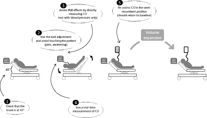
Worth mentioning, though it should be obvious, PLR demonstrates how ridiculous the practice of Trendelenberg Position for resuscitation
Fluid Challenge
500 ml crystalloid or colloid
10-Second Mini-Fluid Challenge
50 ml bolus over 10 seconds through a central line (Critical Care 2014;18:R108) change in VTI measured immediately afterwards
Then Measure the Response
Can Changes in MAP Predict Fluid Responsiveness?
[cite source='pubmed']22278593[/cite], [cite]20111858[/cite], [cite]22464162[/cite]
Most recent analysis states changes in MAP don't predict CI increase from fluid load in septic shock (Intensive Care Med (2012) 38:422–428)
The Cardiac Output Monitors
Marik's Comprehensive Review Article
and my buddy Seth Manoach wrote a nice review as well [cite source='pubmed']22537573[/cite]
NICOM – Bioreactance
- Marik studied 34 patients in the ICU with PLR, NICOM, SVV, and Carotid Flow (The use of NICOM (Bioreactance) and Carotid Doppler to determine volume responsiveness and blood flow redistribution following passive leg raising in hemodynamically unstable patients (Chest 2012 Marik et al.)
- Big validation study showed good accuracy (Intensive Care Med (2007) 33:1191–1194)
- There were a couple of small studies indicating inaccuracy, but when I looked into these–the authors may have had some conflicts
USCOM – Aorta Ultrasound
- Anaesthesia. 2012 Nov;67(11):1266-71.
PiCCO
- PCA + thermodilution
Pulse Contour Analysis Alone
- Bunch of studies keep going back and forth in the lit. I'm not sure if these track changes in afterload. They don't accurately track pressors/inopressors (Anesth Analg. 2011 Oct;113(4):751-7.)
ETCO2
- A PLR-induced increase in EtCO2 >5 % predicted a fluid induced increase in CI >15 % with sensitivity of 71 % (95 % confidence interval: 48–89 %) and specificity of 100 (82–100) %. (Intensive Care Med (2013) 39:93–100)
- Passive leg raise to etco2 (CCM 2014;42:1585)
Carotid and Brachial Artery Analyses
- Search for the evidence on pubmed, it is emerging now
LVOT velocity time integral (VTI)
- Accurate in the hands of experts–kind of annoying to obtain
Can the LV use it?
Dynamic changes can be false if there is RV dysfunction (will yield false positives) as the PPV means the LA needs more fluid but giving it in the face of the RV failure will not get the fluid to the LA. (CCM 2009;37(9):2642)
Pulse Pressure and Stroke Volume Variation
- Predicting fluid responsiveness in the OR (Br J Anaes 2007;98(4):545)
- Editorial on pulse pressure variation
- Original PPV Study
- Dynamic Arterial Waveform Changes[cite source='pubmed']19602972[/cite]
Proposed checklist before performing volume expansion suggested by PPV
[cite source='doi']10.1186/cc13109[/cite]
- Is the patient ventilated with IPPV without spontaneous efforts?
- Is the patient ventilated in non-protective ventilation (tidal volume at least 8 ml/kg)? (EMCrit says 10)
- Is the patient in sinus rhythm?
- Is chest wall compliance normal (thorax closed, no flail chest, no binding)?
- Is the patient unaffected by valvular disease?
- Is the patient unaffected by right ventricle and/or left ventricle dysfunction (for example, as assessed by an echocardiographic examination including measurement of peak systolic velocity of tricuspid annular motion assessed by tissue Doppler echocardiography)?
- Does the patient have normal abdominal pressure?
- Have you decided or have you performed a calibration to decide which threshold value (6 to 20.5%) should be used for the binary decision of volume responsiveness?
- Have you established the compliance of the patient’s vascular capacitance in order to standardize the VE?
- Have you established that the patient’s heart rate/respiratory rate ratio is 3.6?
- Can you safely establish a baseline, perform a volume challenge and remeasure without any factors affecting heart efficiency and/or vasomotor tone during the assessment?
When the percentage of patients who would meet these criteria were evaluated in an ICU, it was achingly small (BJA 2014;112(4):681)
More
- A Critical Challenge to the accuracy of SVV during mech vent both for absolute and as a trend (Crit Care Med 2012;40:1186)
- Put the patient on 10 ml/kg and sedate them. (CCM 2009;37(9):2642) Driving pressure > 20 is probably necessary to get good PPV (Inten Care Med Volume 36, Number 3 / March, 2010:1432)
- Slow the arterial line trace to 6.25mm/second to synch with the capnograph. Makes it easy to identify systolic pressure variation (twitter anesthesia tips)
Pleth Index
- anesthesia study
Can the RV take it?
IVC Ultrasound
I was just thinking about it the wrong way
CVP
I think this is the only ? CVP may be able to indicate, and probably not as well as IVC
Other Stuff
Easy-Peezy for Quick ?s of Squeezy
End-Expiratory Occlusion Test
(Crit Care Med. 2012;40:152–157)
15 sec end-exp occlusion, look for pulse pressure >=5% during the end-expiratory occlusion with a sensitivity and a specificity of 87% and 100%, respectively, and by an increase in cardiac index >=5% during the end-expiratory occlusion with a sensitivity and a specificity of 91% and 100%, respectively
Works even with high PEEPS (Crit Care Med 2013;41:1692)
End-exp occlusion was good in pts with poor resp compliance; PPV was not (Crit Care Med 2012;40(1):151)
Expiratory Hold to Predict Volume Responsiveness
(Crit Care Med 2009;37(3):951)
15 second expiratory hold, maximal change during last five seconds=test. If the patient is spont breathing, change the trigger settings to make it hard to take a breath. 5% change in arterial pulse pressure predicts volume expansion as does increase in CI.
And even more controversies and thoughts
In Critical Care Medicine, an editorial by Hilton and Bellomo questioning whether fluids should be given in sepsis in any amount greater than a couple of liters; they want to know where the evidence is…
Another from Paul Marik and another from Paul Marik
An article seems to indicate that the best predictor for development of pulmonary edema during fluid loading is reaching plateau of CI, indicated by no additional increase with fluids (CCM 2012; 40:793) as shown graphically by the Marik-Phillips Curves


See this great Meta
What do you think? Comment below
Additional New Information
More on EMCrit
- Podcast 86 – IVC Ultrasound for Fluid Tolerance in Spontaneously Breathing Patients – EAT IT STONE(Opens in a new browser tab)
- SMACC Debate – Rob MacSweeney vs Paul Marik – Predicting Fluid Responsiveness is a Waste of Time
- Podcast 112 – A Response to the Marik Sepsis Fluids Lecture
- Podcast 345 – CLOVERS Trial and Fluids in Sepsis
Additional Resources
You Need an EMCrit Membership to see this content. Login here if you already have one.
Professor
Nassau University Medical Center
No conflicts of interest (coi).
- EMCrit 373 – Mike Weinstock with another Critical Care Bounceback: “Asymptomatic Hypertension” - April 18, 2024
- EMCrit Wee – Ross Prager on 10 Heuristics for the New ICU Attending - April 13, 2024
- EMCrit 372 – FoundStab Intubation SOP - April 5, 2024
I’ve been wondering. What is it about IVC us imaging that makes it better than CVP monitoring? It would seem the disadvantages of CVP would be the same as with IVC monitoring
IVC as we teach it is the dynamic assessment of size in response to respiratory variations (primarily in spont breathing patients) as opposed to static CVP
So, to use an example, I’ve had multiple post abdominal surgery patients to care for as of late. Patients in whom because of dressings, because of discomfort, and because of my somewhat amateur ultrasound skills, I am unable to obtain us of their IVC. I’m still looking for objective assessments of volume status for these patients, instead of the surgical mantra of “low urine output = give more fluids”. What I’ve been turning to is CVP monitoring using a transducer on a supra diaphragmatic CVC. Not for looking at the actual number mind you, I don’t really care about the… Read more »
We can imagine that the IVC-diameter respiratory variations reflect the respiratory changes in CVP and, as it was elegantly explained by Magder ( ….Guyton at the bedside Critical Care 2012, 16:236), these changes can be indicative of good response to fluids but not necessarily. I imagine that if the IVC empties itself in inspiration this means that the right ventricle is able to pump forward the bigger amount of blood received but we can’t be sure that also the left ventricle is able to do the same. In my experience I’ve seen and documented more then one patient in overt… Read more »
Thanks for another great post. I’ve found this is a very difficult topic in the ICU, where frequently we have patients who are still on pressors after the initial resuscitation and there is an almost insatiable desire to give more fluid in order to get the pressors off as quickly as possible. Rarely is it asked whether this will work or will benefit the patient. Indeed, it can be difficult to extubate a patient who is 10 liters positive over 24 hours after their vasoplegia has resolved. I think there is a population of patients who are temporarily fluid responsive… Read more »
I have stood by and watched so many differently trained folks battle this out for more than a decade of my life. I can not for the life of me find any study, any methodology, or dogma that puts me any closer to what works. I have seen stuff come and go…cvp, leg raise test, p to p variation in a line tracing, SWANS, and bedside echo. After all the academic discussion and articles written, we come back to the ever so humbling patient’s physiology that particular day. I have been ever humbled about how much I do not know.… Read more »
Wonderful hearing about the EGLS. I do not feel we can do justice to the septic patients without an echo. It is wonderful hearing about the right ventricle. As EP’s we seem to like the sexy stuff i.e. LV, Systemic BP and Systolic function but in general the not-so-sexy stuff like RV, Pulmonary circulation and Diastolic function is often ignored. In my practice in a non tertiary ED, I am seeing quite a few patients with Pulmonary hypertension, quite often Type 3 undiagnosed. Even those patients where you are suspecting diastolic dysfunction with your E/A ratios, the essence will be… Read more »
Ultrasound is a tool which has limitations as Scott nicely pointed out. PA catheters are safe despite what most people think and the key to its use is proper patient selection which the trials did not do. In the patient with established baseline cardiomyopathy, or the patient with mixed shock or undifferentiated shock I have seen even the best ultrasonographers in the nation scratch their heads because bedside TTE is limited, also if you have poor windows which many of our pts have you’re just guessing. We need to bring back the PA catheter because it can be useful in… Read more »
you talk about an increase of 5% in EtCO2 as predicting responsiveness. Does this mean if the etco2 is 30 that it would have to go up by 1.5 to be significant?
How soon after a PLR do you measure the new C.O?
yep, 5% was the study parameters. Spec of 100% and sens of 71% (so believe a positive, not a negative) based on a small study.
they saw the increase during the 1st minute after PLR maneuver.
Although tricky to perform sometimes, the SV measurement from the VTI/LVOT method (and subsequently the SV response to fluid challenge) seems the most accurate to me (mathematically). Any data or support for that?
Arnav
medicine resident at columbia prebyterian
I think that, in this setting, wondering if the patient is fluid responder or not isn’t the right question. In my opinion what’s really matter is to understand if the patient requires to increase his CO and consequently his DO2 or if he needs to increase his SVR and consequently his BP. If I can know since the beginning that my patient has a CI of 4 or more, which is the benefit in increasing his CI to 5 or more? Marik has teached us that the so called oxygen debt in sepsis probably doesn’t exists. Lactate probably is expression… Read more »
Why isn’t anyone talking about Swans?! Especially the more senior docs? While there is something to be said for technology and non invasive methods of assessing your patient, we can’t neglect the fact that in some sub groups, invasive means have a role! In septic patients with arrhythmias (afib) and/or heart failure, septic cardiac hypokinesis, or significant pulmonary hypertension (primary and even sometimes secondary) I have a newfound love for Swans. While bedside echo is great, it’s too labor intensive for me. I cover 20 ICU beds and I’m the emergency surgery at night with just residents (vast majority of… Read more »
Hello Scott thanks for your fantastic podcasts. Im last year medical student in Vienna, Austria, as well as paramedic and PhD engineer, and am very interested in emergency medicine and critical care. I (think I) can grasp most of what you have talked about, but even if it might be a silly question, I wanted to discuss with you and the audience the following: the approach to volume assessment by asking the questions -can the RV take it, can the LV use it- seems very logical, but what about ‘can the LV take it’? I was wondering, is there no… Read more »
LV failure very rapidly translates into RV increased pressure, and B-lines on ultrasound. The answer to can the RV take it is the answer to can the LV take it
Fantastic and comprehensive overview Scott, as always. One thing I would like to highlight, and which in part may explain many of the shortcomings of the IVC ultrasound in the literature, is the manner in which it is measured, which is in diameter at an arbitrary level. The physiology we are assessing when looking at the IVC is that of volume variation. It is exceedingly difficult to assess a volume when using a linear measurement… Hence the importance of looking at a significant portion of the IVC both in long and short axis. In effect, this is “eyeballing” the IVC,… Read more »
Wonderful! Your comments regarding the fact that volume overload and volume responsiveness may overlap were game changing: everybody should read this post: http://thinkingcriticalcare.com/2016/02/21/volume-responsiveness-and-volume-tolerance-a-conceptual-diagram-foamed-foamcc-foamus/
Much appreciated Scott! Could make for a good RLA lecture!
cheers!
Phil
[…] https://emcrit.org/emcrit/assessing-fluid-responsiveness/ […]