

(Contains cutting-edge ideas, some of which lack evidentiary support: caveat emptor; more on the bleeding edge series here)
Scott Weingart just posted a podcast about management of the hypercapneic, obtunded COPD patient who is failing BiPAP. Do we need to intubate these patients, or could we somehow clear their CO2 noninvasively? This post will start off by exploring respiratory drive as a mediator of disease. With that groundwork, we'll explore the obtunded COPD patient.
Getting started: a graphic representation of respiratory drive
Minute ventilation is related to the work of breathing and the PaCO2 as shown below (1). The respiratory drive has a modulating effect on the entire system:
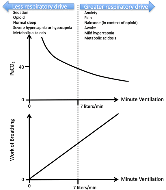 Normally, this system is in under tight control. For example, if you hold your breath, your PaCO2 starts to rise gradually. Mild, acute hypercapnia causes a profound increase in your respiratory drive – forcing you to start breathing.
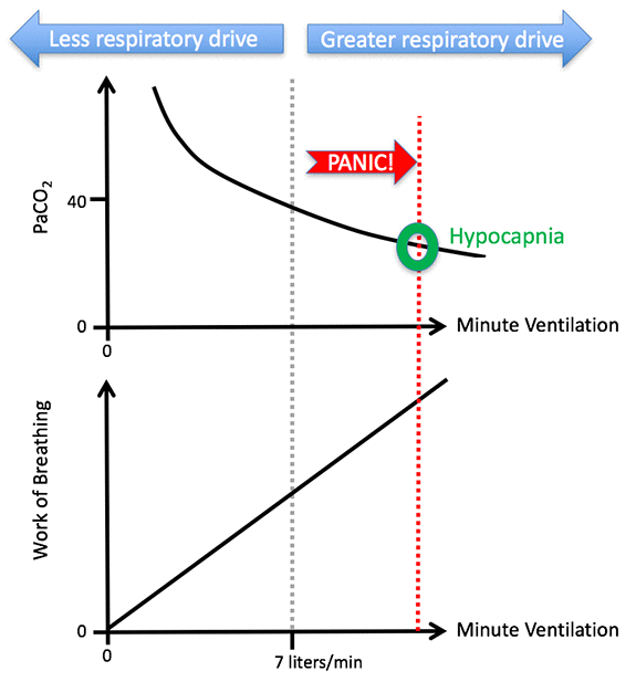
A panic attack illustrates how things can go awry. Normally, mild hyperventilation causes hypocapnia, which reduces the respiratory drive and brings the system back to normal. However, during a panic attack, severe hyperventilation causes alkalosis and hypocalcemia, which in turn triggers worsening anxiety. This further increases the respiratory drive, causing further hyperventilation – a vicious cycle that spirals out of control:
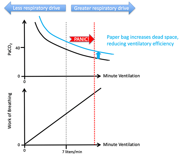
 This cycle can be broken by using a paper bag to cause rebreathing carbon dioxide:
 Respiratory drive is involved in two life-threats.
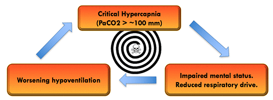 The more obvious threat is critical hypercapnia, because this may eventually lead to a comatose state with inability to protect the airway, apnea, and death. Beyond a certain point, hypercapnia becomes self-perpetuating (because severe hypercapnia has an anesthetic effect which decreases the respiratory drive). This is widely feared, but in practice it generally isn't an immediate life-threat among hospitalized patients (it usually occurs gradually and is easily fixed with noninvasive or invasive ventilation).
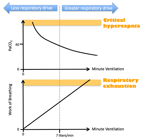 The less obvious life threat is excessive respiratory drive which eventually precipitates diaphragmatic exhaustion. This is probably the more common scenario for respiratory arrest in a hospital. It begins with an increase in the work of breathing (e.g. from pneumonia). Over several hours the patient is tachypneic in a compensated state, with gradually deteriorating diaphragmatic strength reserves. Eventually, respiratory muscles become exhausted and the patient is unable to cough and clear secretions. Mucus plugging or complete exhaustion occurs, precipitating very rapid deterioration and respiratory arrest (2).
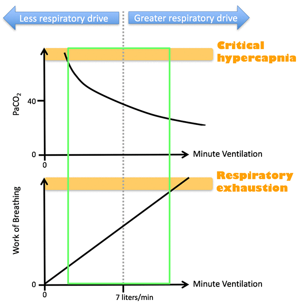
Range of safe minute ventilation (green box of goodness)
These two life-threats define a range of minute ventilation which is compatible with survival (green box below). A minute ventilation below this range poses a threat of critical hypercapnia. A minute ventilation above this range creates a threat of respiratory exhaustion:
 Applications
The following scenarios illustrate how this physiology plays out in clinical medicine. They build upon one another with escalating complexity.
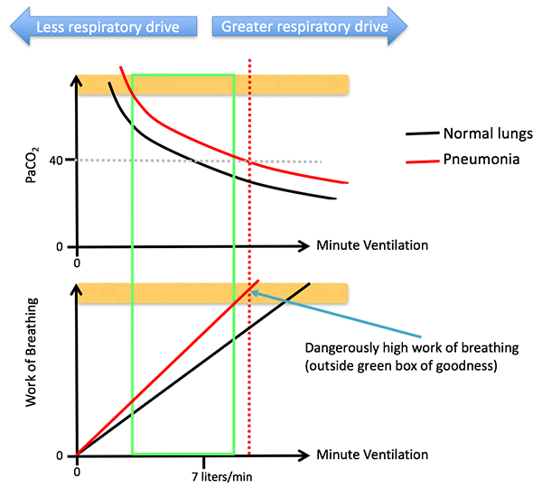
Application #1: Using high-flow nasal cannula for pneumonia
Let's start with the pneumonia patient, who has these problems:
- Impaired CO2 clearance, shifting the PaCO2 curve upwards.
- Reduced lung compliance, increasing the work of breathing.
The patient's brain is still trying to achieve a normal PaCO2. To achieve this, the minute ventilation must be increased substantially. However, this requires a dangerously high work of breathing, placing the patient at risk of exhaustion:
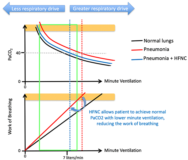
 High Flow Nasal Cannula (HFNC) reduces the anatomic dead space, improving the efficiency of ventilation (explained further here). This shifts the PaCO2 curve downward, back towards normal. The patient can now achieve a normal PaCO2 with a lower minute ventilation, which requires less work:
 Application #2: Opioid for acute asthma
 Asthmatics often fall into a vicious cycle involving respiratory failure and tachypnea. As we know from invasive mechanical ventilation, asthmatics fall apart when they are tachypneic (this causes inadequate expiratory time, leading to AutoPEEP). In a non-intubated patient, AutoPEEP cannot be easily measured. Nonetheless, it is hugely problematic because it causes hyperinflation (which compromises diaphragmatic function) and makes it difficult to inhale (patients must overcome the AutoPEEP to draw air into the chest).
This may be diagramed as shown below. The work of breathing curve is curvilinear, because work increases exponentially at higher minute ventilation (due to tachypnea-induced AutoPEEP).
 My usual treatment for this involves BiPAP and dexmedetomidine, to support the work of breathing and reduce anxiety (discussed further here). However, another possible therapy is opioid. This could break the cycle as shown here:
 The effect of opioid is diagrammed as below. Opioid would drop the respiratory rate to a reasonable level, reducing AutoPEEP. It might even induce a mild (safe) amount of hypercapnia. The key is that opioid would reduce the work of breathing to a sustainable level, avoiding respiratory exhaustion.
 I haven't needed to use this, and it remains unproven. However, some intensivists have reported success with it (Stemp 2013). This could be accomplished as follows (3):
- Opioid must be started early, before the patient develops complete respiratory exhaustion.
- Small divided doses of IV fentanyl are ideal (due to rapid onset). Titrate fentanyl against respiratory rate, with a goal of dropping the respiratory rate to ~15-20 breaths/minute.
- Avoid benzodiazepines. Benzodiazepine reduces the margin of safety with opioids, without directly treating the underlying problem (tachypnea).
- Intensive monitoring is required. Ideally these patients would be on BiPAP, allowing monitoring of respiratory rate and tidal volume.
Application #3: Obesity hypoventilation syndrome
Morbid obesity increases the work of breathing because the diaphragm must push against the abdominal contents. This leads to a situation where, in order to achieve a normal PaCO2, an excessively large work of breathing would be required:
 This scenario would be rapidly fatal. To avoid death, the brain adapts to morbid obesity by targeting a higher PaCO2 level. Mild-moderate hypoventilation reduces the work of breathing to a tolerable level:
 Application #4: COPD phenotypes: Blue Bloaters vs. Pink Puffers

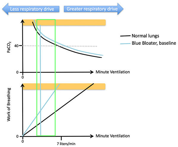
 The two most stereotypical forms of COPD are pink puffers and blue bloaters, as shown above (4). These patients have different physiology and clinical presentation:
- Pink Puffers: These patients have classic “dry emphysema,” with destruction of the alveoli causing ineffective CO2 clearance and airway obstruction. Patients are trying to maintain a near-normal PaCO2, causing chronic dyspnea (eventually leading to weight loss from persistently increased work of breathing). Overall this physiological derangement is similar to asthma as discussed above. The primary life-threat is increased work of breathing leading to diaphragmatic exhaustion. These patients present with respiratory distress and tachypnea.
- Blue Bloaters: These patients have an overlap syndrome of COPD plus obesity-hypoventilation syndrome. The combination of obesity plus airflow obstruction causes a dramatic increase in the work of breathing, which is the dominant physiologic problem. To adapt, patients develop substantial chronic hypercarbia. When these patients decompensate, they manifest with severe hypercapnia, hypoventilation, and obtundation.
 Application 5: Blue Bloater with critical hypercapnia

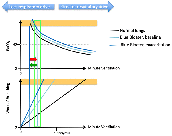
Let's delve a bit deeper into Blue Bloater physiology. At baseline, these patients have markedly increased work of breathing and mildly impaired ventilation. They compensate by living with chronic hypercapnia, which avoids diaphragmatic fatigue.
 During an exacerbation, bronchospasm causes increased work of breathing and also impaired CO2 clearance. This causes the patient to become extremely unstable, with a very narrow range of safe tidal volumes (narrow green box, see below). Eventually the patient develops some muscle fatigue, the PaCO2 increases, and the patient develops critical hypercapnia. This causes progressive mental status decline, reduced respiratory drive, and a spiral of hypoventilation.
 The first treatment that we apply is BiPAP. BiPAP reduces the work of breathing by providing mechanical support, as shown here:
 The problem is that if the patient is too hypercapneic, they may continue to remain in a stuporous state with poor ventilatory drive, even despite the BiPAP. The patient has fallen off the left side of the curve, and may tend to remain there. Such patients have typically required intubation to control their hypercapnia.
Finally… avoiding intubation in the critically hypercapneic COPD patient
Now let's consider the critically hypercapneic COPD patient who doesn't respond immediately to BiPAP. Is there some way to avoid intubating this patient?
Beware of transient improvement
It's tempting to think that if we could ventilate the patient for several minutes and blow off their CO2 (e.g. with bag-mask ventilation), they would wake up and recover. However, short-term ventilation alone may be insufficient. The risk here is that the patient will improve (red arrow below), but later on will deteriorate and slip off the curve again (green arrow):
 The classic example of this phenomenon is the patient with difficulty tolerating BiPAP who presents obtunded with critical hypercapnia. BiPAP is initiated, causing the patient to blow off CO2 and gradually wake up. After the patient wakes up, he rips off the BiPAP mask. A few hours later, CO2 builds up again and the patient returns to his prior obtunded, hypercapneic state. The patient is now obtunded enough to tolerate BiPAP, BiPAP is re-started, and the entire cycle repeats itself. This cycle could be resolved by using BiPAP and then starting dexmedetomidine when the patient wakes up (to promote mask tolerance without suppressing respiration). After ~24 hours of diaphragmatic rest and bronchodilation, the patient may be able to get off BiPAP and stay off.
I've seen similar cases where a patient presents to the Emergency Department obtunded, and responds well to BiPAP. Upon arrival in the ICU, the patient looks fantastic. BiPAP is discontinued. Within a few hours, the patient relapses into an obtunded, hypercapneic state.
The reason that a single episode of hyperventilation isn't sufficient is that these patients are suffering from more than just hypoventilation. The underlying problems often include exacerbation of bronchospasm and diaphragmatic fatigue. Fixing the PaCO2 doesn't fix all of these problems, leaving patients at risk for relapse.
BiPAP sometimes works if given for longer periods of time
I've encountered several hypercapneic obtunded COPD patients who failed to respond to BiPAP over a few hours and were DNI (Do Not Intubate). These are patients who would definitely have been intubated, if not for their DNI status. Following discussion with their families, I will frequently offer an extended trial of BiPAP (5).
These patients usually do surprisingly well. Upon admission they are obtunded and look pretty terrible. Overnight they are treated with steroid, antibiotic, bronchodilator, and high BiPAP settings (e.g. ~20cm/5cm). They sleep deeply, hypoventilate, and rest their respiratory muscles. Their bronchi open up gradually. The next morning they will wake up and demand breakfast.
Thus, there is definitely some population of patients who don't respond immediately to BiPAP, but who will respond to an extended trial of BiPAP (plus steroid/antibiotic/bronchodilator). This is supported by literature showing that many patients who are DNR/DNI and treated with BiPAP will survive. The weakness of this strategy is that it leaves patients in an obtunded state for a while, theoretically increasing the risk of aspiration (6).
Controlled Burn: Induction therapy to facilitate CO2 removal and stabilization
Controlled burn refers to a strategy to rescue patients from the spiral of increasing hypercapnia and somnolence, without intubation. One could imagine roughly two ways to do this, as shown below. The first approach (“anesthesiology approach”) is to start with a mechanical strategy to increase minute ventilation (e.g. bag-mask ventilation or laryngeal mask airway ventilation with propofol). Once the PaCO2 was decreased enough to improve the patient's consciousness and respiratory drive, they could be placed on BiPAP as a maintenance therapy to stabilize them and prevent recurrence.
 I've never specifically tried to do this. However, one possible strategy might be as follows (7):
- Drop a nasogastric tube and place it to wall suction to prevent gastric insufflation. Perform bedside abdominal ultrasound to confirm that the stomach is decompressed (8).
- Initiate full-face BiPAP with very high settings (e.g. 30 cm iPAP / 5 cm ePAP). Set a backup rate of ~14 breaths/minute with an inspiratory time of 1 second. Generally such aggressive BiPAP settings are impossible because the patient wouldn't tolerate them and gastric insufflation could occur. However, in a somnolent patient with a nasogastric tube they could be reasonable.
- Ensure an open airway (e.g. by placing patient in sniffing position, and possibly using a nasopharyngeal airway).
- Monitor patient intensely for emesis, intolerance, or hypoventilation. Monitor tidal volumes and minute ventilation on the BiPAP machine. If the tidal volume and minute volume are low, this is an early indication that this strategy will fail. However, if tidal volumes and minute ventilation are good, this suggests that you might not get much added benefit from invasive ventilation (aside from airway protection of course)(9).
- Ideally the patient would start waking up and breathing more, allowing you to gradually scale back the BiPAP pressure and transition to a more traditional BiPAP setting. If the patient woke up and became agitated, dexmedetomidine may be considered to control agitation without compromising respiratory drive or airway-protective reflexes.
 An alternative strategy is “medical induction” therapy. In this strategy, the patient would be placed on BiPAP at high settings, but within the range of conventional treatment (e.g. 20 cm iPAP / 5 cm ePAP). Medical therapy would be provided to stimulate the respiratory drive. Depending on the scenario, one or more of the following treatments could be used:
- For a patient on chronic opioids, tiny doses of titrated naloxone will cause a substantial increase in respiratory drive. Be careful not to induce withdrawal, however (which could be dangerous, particularly if it causes vomiting).
- Mechanical or auditory stimulation (e.g. someone at bedside who keeps on stimulating the patient to prevent falling asleep).
- Intravenous aminophylline. This causes bronchodilation and stimulates the respiratory drive. It has been proven to reduce PaCO2 in an RCT involving patients with COPD exacerbation (Duffy 2005). Use may be limited by a side-effects including nausea and vomiting.
- Intravenous caffeine. Caffeine has a similar mechanism of action and respiratory effects compared to aminophylline (Evans 2017). It is often used to stimulate breathing in neonatology (Dobson 2016). One RCT showed that 500 mg IV caffeine benzoate reduced post-operative respiratory complications among a group of 60 patients with sleep apnea (Gouda 2010). Compared to aminophylline, caffeine seems safer and easier to use (single dose of caffeine versus bolus plus infusion with aminophylline).
- Intravenous acetazolamide and/or normal saline infusion for patients with severe iatrogenic metabolic alkalosis (e.g. due to over-diuresis)(10). In this situation, gentle reduction in bicarbonate may increase respiratory drive. However, be careful that you don't reduce the bicarbonate below the patient's baseline – this could increase respiratory drive excessively.

- Respiratory drive is a key mediator of disease, making it a potential target for intervention.
- Respiratory drive can create two potential life-threats. Grossly inadequate drive may cause hypercapnia and coma. Excessive drive may cause respiratory exhaustion leading to respiratory arrest.
- Many factors affect respiratory drive (e.g. anxiety, pain, opioids, naloxone, sleep, wakefulness, metabolic acidosis/alkalosis, caffeine).
- Artful manipulation of respiratory drive may stabilize patients with respiratory failure. This could be useful in conditions with severe tachypnea (e.g. asthma) and also in conditions with critical hypoventilation (e.g. COPD with hypercapneic encephalopathy).
Related
- Controlled burn for hypercapneic encephalopathy in COPD (EMCrit podcast 213)
- Opiates for severe asthma: Google-plus conversation between Leo Stemp and Reuben Strayer.
- Dexmedetomidine to facilitate noninvasive ventilation (PulmCrit) – some discussion here about the role of anxiety and tachypnea in causing respiratory deterioration in COPD/asthma.
- Apneic ventilation using pressure-limited ventilation (PulmCrit) – discussion of using a V60 BiPAP to provide apneic ventilation prior to intubation. A similar strategy could be used for the controlled burn.
Notes
- Seven liters/minute is used arbitrarily as a normal minute ventilation at rest, but this will vary depending on the patient's size and metabolic activity.
- Hypercapnia does occur in this scenario, but only in the moments immediately preceding death. Thus, using an ABG to “screen for respiratory failure” in this scenario is worthless. The ABG will look unremarkable (and falsely reassuring) until the patient is coding.
- This is based on Dr. Stemp's description which can be found in attachments here.
- I apologize if anyone is offended by this terminology, but it is standard verbiage and also fairly descriptive. This is a tricky topic, so any terminology which helps us understand it will ultimately help us take better care of our patients and thus be in their best interest.
- A long time ago, I used to be frustrated by DNR/DNI patients in the ICU, since I felt helpless to treat them. However, I've realized that there is often quite a lot that we can do for these folks. Once conventional therapy is failing, it becomes reasonable to try outside-the-box therapies (with consent). For example, I've encountered several elderly patients with septic shock who were DNR/DNI and refused central line – these patients are very often curable with peripheral pressors, steroid, conservative fluid strategy, ascorbate, ephedrine PRN, etc.
- It is unknown how the risk of aspiration compares to the risk of ventilator-associated pneumonia. The risk of ventilator-associated pneumonia is about 2-3% per day initially, so intubating a COPD patient probably carries about a 5% risk of ventilator-associated pneumonia. It's possible that the risk of BiPAP-associated aspiration is less than this risk of ventilator-associated pneumonia – unfortunately I'm unaware of any solid data on this.
- There are lots of ways you could do this: LMA plus propofol, bag-valve-mask, volume-cycled noninvasive ventilation, etc. I've described an approach using pressure-limited BiPAP because that's usually my weapon of choice, but you could use anything that you're good at. The key is monitoring it and seeing that it works.
- It should be possible to achieve a reasonable mask seal despite the NG tube.
- Note however that if there is significant mask leak, the BiPAP machine will over-estimate tidal volume and minute ventilation. It may also useful to physically examine the patient to see the amount of chest rise and air movement that is being achieved.
- Metabolic alkalosis is one of the few situations where it's a physiologically logical move to use normal saline. Saline is an acidotic fluid, so it helps improve the alkalosis.

- Pulmcrit wee: The cutoff razor - April 15, 2024
- PulmCrit Blogitorial – Use of ECGs for management of (sub)massive PE - March 24, 2024
- PulmCrit Wee: Propofol induced eyelid opening apraxia – the struggle is real - March 20, 2024
I’ve used opioids before for patients who remain panicky and tachypneic on NIV. Their “air hunger” seems to outstrip what the machine can give them. It seemed like a simple decision to me – the only major risk would be respiratory depression, which might in the worst case necessitate intubation. But we’re talking about patients you’d have to intubate anyway if you couldn’t take away that air hunger, as they keep ripping off the mask and exhausting themselves.
This brings us to the topic of how to sedate patients to facilitate tolerance of noninvasive ventilation – a topic which could easily occupy a full blog or podcast by itself. Not much is known about this (e.g. review article by Longrois 2014 https://www.ncbi.nlm.nih.gov/pubmed/25699177). It’s very hard to say anything about this topic with certainty, but here are some thoughts – – Dexmedetomidine is generally my go-to drug here on the basis of safety and titratability. Drawbacks include cost and slow onset. With the exception of bradycardia/hypotension, this brings a lot of goodness with little risk. – Opioids (esp titrated… Read more »
fantastic post and such a wonderful foundation to bolster my last podcast. No love for ketamine for asthmatics? Much quicker to kick in than dex and no resp depression like opioids.
Thanks much. Great question, I don’t know the right answer but this is how I’d play it. I think the key question is how sick the asthmatic is, specifically if they look like they might need to get intubated within the next 15-30 minutes: <1> If they are super-sick and look about to crash, then ketamine is probably the way to go. As you pointed out, this gets immediate behavioral control and bronchodilation. Fastest option. <2> If they are sick but look like they can wait 20-30 minutes, then dexmedetomidine +/- a touch of fentanyl might be a reasonable way… Read more »
What about Ketamine infusion? I’ve had patient’s that I couldn’t “chill out” with Dexmedetomidine, and the idea of a ketamine load and infusion seems like it could be interesting, especially in the asthmatic.
Application #3: “To avoid death, the brain adapts to morbid obesity by targeting a _lower_ PaCO2 level.”
Shouldn’t it say “a _higher_ PaCO2 level”?
you are entirely correct, I’ve fixed the blog. thanks much.
post-publication error detection in 4 hours.
I’ve had some anecdotal success (although I have yet to be able to figure out why) with AVAPS (via the Respironics V60) when these hypercapnec COPD patients seem to be failing conventional bipap settings. I don’t know if it is a result of the same phenomenon as you discussed with just happening to give more time for recovery, or if AVAPS is truly providing more efficient ventilation. It’s usually a salvage therapy as a last resort before intubation, and have had some patients do surprisingly well. Any thoughts?
Excellent point. If the patient is on BiPAP but their intrinsic respiratory rate is low, adding a backup rate could help improve minute ventilation. Anything that increases minute ventilation will work in your favor.
Also possible that a very mild level of ventilator dyssynchrony created by introducing a backup rate could stimulate the patient and actually cause them to breathe more (acting as a very mild aversive stimulus that prevents them from falling asleep).
What’s your go to starting dose of fentanyl for this purpose, assuming an opiate naiive 70 kg male? 25 mcg- 50 mcg? How about very low dose naloxone in chronic opiate dependent patients?
1) Haven’t done this but would guess 25 mcg might be reasonable. It works rapidly so you could titrate with repeated doses. 2) Yes, definitely. This works but you need to be very gentle with naloxone (initial dose of 0.1 mg or less). Once you titrate up to a dose that keeps the patient breathing but not in withdrawal an infusion will generally be needed, the infusion dose can be started at half of the effective dose per hour (e.g. if the patient needs 0.2 mg initially then drip 0.1 mg/hr). Obviously ongoing close monitoring required with this to avoid… Read more »
Thanks! Another thing I was wondering was what amount of CPAP pressure to use for asthma? I’m sure you would need more in an obese patient, but in a patient with a normal body habitus, would 8-10 cm H2O be a reasonable place to start?
A little PEEP might help stent open the airways during exhalation, but too much PEEP can impair exhalation (reduces the driving pressure). People have strong opinions about this but I’m not aware of any solid data. 5-8 cm is probably reasonable.
Is there an estimated rate of decrease of respiratory rate in relation to a certain dose of narcotics? For instance for every 25mcq of fentanyl rate will decrease by 2-5. Hopefully that makes sense.
So far as I can tell it’s highly unpredictable. Patients with a history of opioid use may be tolerant. The nice thing about fentanyl is that it works fairly rapidly, so you can give small doses and frequently assess how the patient is responding.
Hi Josh Thanks for this great post. I came across the scenario 2 recently in an asthmatic with hyperventilation from anxiety/panic. Her wheeze had improved but her chest remained tight. I used a dissociative dose of ketamine, to bronchodilate and to “turn off” her cerebral cortices. This did cause her to become apnoeic and needed bagging with an OP airway. Her chest didn’t rise initially and she desaturated but with good two person technique, we were able to bag her up & her respiratory drive kicked in. I think this happened because her PaCO2 was low to begin with so… Read more »
it sounds like the patient somehow received a bolus of ketamine. A bolus of dissociative-dose ketamine will often cause apnea for 60-90 seconds. If ketamine is given as a slow IV push over about two minutes this won’t happen. Have had the same thing happen to me (likewise the patient was fine but it scared us). Unfortunately ketamine is often touted as causing no respiratory suppression but this warning should be somewhere in the fine print.
Hi,
The quality of your blogs are so amazing, no wonders I keep coming to your website
to read more such blogs.
Great work….!
Source: https://tractorguru.in/mini-tractors-in-india
Trying to reduce TcCO2 levels in a patient with severe restrictive lung disease, bilateral pleural plaques, pulmonary hypertension and progressive SOB. His latest ABG showed blood pH 7.37, PaCO2 62 and Bicarbs 35. I’m trying to flush out some of that CO2 with BiPAP but no luck. His TcCO2 reading is 77 during sleep and he started to hyperventilate when I raised the BiPAP delta. Not sure what steps to take with regard to PAP – I tried AVAPs, but ended up switching him back to a straight CPAP as he began to hyperventilate (BR 40) and after an hour… Read more »
My clinical question regards focusing on the respiratory drive as opposed to the underlying obstruction in asthmatics. Logically, an asthmatic is hyperventilating as the chemosensitive centers respond to blood-gas/acid disturbances. Therefore, why would one administer an opioid to reduce the respiratory drive in a crashing asthmatic (with the hope of allowing for diaphragmatic rest) as opposed to treating the dynamic hyperinflation extending from severe obstruction? Why not just administer intravenous epinephrine and potentially theophylline if inhaled bronchodilators fail? I could only understand using opioids once the oxygen saturation has normalized and the patient still remains tachypneic. However, I could not… Read more »
Great job on writing such an insightful and informative article! Your attention to detail and thorough research really shines through in your writing. I appreciate how you presented the information in a clear and organized manner, making it easy for readers to understand and follow along.
<a href=”https://trucks.tractorjunction.com/en/mahindra”>Mahindra truck price</a>